흉선종 재발과 중증근무력증 악화 사이의 시간적 연관성
Temporal Association between Thymoma Recurrence and Exacerbation of Myasthenia Gravis
Article information
Trans Abstract
Background
This study investigates the potential link between myasthenia gravis (MG) exacerbation and thymoma recurrence in patients with thymoma-associated MG (TAMG).
Methods
We conducted a retrospective medical record analysis at Severance Hospital. Baseline clinical feature of MG, initial histological features, and severity of MG were recorded. Based on the change in MG activities of daily living (ADL) score and MG-spsecific treatments, patients' MG status was assessed over the 2-year period around thymoma recurrence, and was classified into three categories: improved, unchanged, and worse.
Results
Of the 178 patients with TAMG, 18 (10.1%) experienced recurrence of thymoma. Mean age at initial thymectomy and at thymoma recurrence was 44.1±9.4 and 48.8±9.1 years, respectively. At the point of thymoma recurrence, myasthenia gravis foundation of America post-intervention status was complete stable remission in four (22.2%), pharmacologic remission in five (27.8%), minimal manifestation in five (27.8%), improved in two (11.1%), and unchanged in two (11.1%) patients. Median MG ADL score was 0.0 (interquartile range [Q1-Q3], 0.0-1.8). During 1-year period before recurrence of thymoma, three patients experienced worsening, probably associated with infection and discontinuation of medication. During the 1-year period after the recurrence, four patients experienced worsening after the second thymectomy.
Conclusion
MG worsened in 16.7% and 22.2% of the patients before and after thymoma recurrence, respectively. Exacerbation of MG in cases of thymoma recurrence seems more likely to result from known risk factors, including infection, general anesthesia, or discontinuation of medication, rather than from the recurrence of thymoma itself.
Introduction
Myasthenia gravis (MG) is an autoimmune neuromuscular disorder in which autoantibodies against the structure of neuromuscular junction induce weakness of ocular, bulbar, limb, and/or respiratory muscles. Approximately 10-20% of patients with MG are known to have thymic neoplasm, most commonly thymoma. Thymomas are neoplasms of thymic epithelial cells. Alteration of thymic microenvironment by the neoplasm may lead to the release of self-reactive T cells to peripheral circulation, causing various paraneoplastic syndrome including MG.1 Individuals with MG and thymoma, a condition known as thymoma-associated MG (TAMG), are known to experience more severe symptoms of MG and likely to have higher levels of acetylcholine receptor antibodies (AChR Abs) than the patients with MG without thymoma.2 Thymectomy is recommended to the patients with TAMG for the management of tumor.
Recurrence of thymoma after the resection of thymoma is not uncommon. The chance of thymoma recurrence after complete resection of the initial thymoma ranges from 5% to 50%.3-5 Recurrence is usually confined to the pleural dissemination or local relapse, while distant metastasis is rare. Pleural recurrence of thymoma is often asymptomatic and is frequently detected incidentally during routine follow-up. When symptomatic, clinical manifestations commonly include pleural effusion, thoracic discomfort, and dyspnea. Interestingly, symptoms of MG can exacerbate along with the recurrence of thymoma. A previous study showed that thymoma recurrences were accompanied by worsening of MG.2 Another study similarly observed that exacerbation of MG can be accompanied by recurrence of thymoma and that many of these recurrences were identified due to the worsening of MG symptoms.6 However, these studies are based on small number of patients and clinical characteristics of MG were not thoroughly evaluated.
In this study, we intended to investigate the potential link between thymoma recurrence and exacerbation of MG in patients with TAMG. Patients with TAMG who experienced the recurrence of thymoma were identified. The proportion of patients with TAMG who experienced the exacerbation of MG during the period one year before and after the recurrence of thymoma was analyzed.
Methods
Patients
This retrospective study investigated the medical records of the patients with TAMG who visited the Severance Hospital between January 2010 and January 2024. Diagnosis of MG was made based on clinical, serological, and electrodiagnostic evaluations. Of the patients with TAMG, the patients 1) who underwent thymectomy at Severance Hospital and 2) whose thymoma was histologically confirmed after thymectomy were included. The patients whose medical records were insufficient to assess the clinical course of MG was excluded. Patients who were finally included were classified into the patients who experienced recurrence of thymoma (recurrence group) and whose who did not (non-recurrence group). The requirement for obtaining informed consent from study subjects was waived by the Institutional Review Board (IRB) of Severance Hospital, Yonsei University College of Medicine, Seoul, Korea (IRB No. 4-2024-1322).
Follow-up after thymectomy
In our institution, patients with thymoma undergo regular follow-up by thoracic surgeons to monitor the recurrence of thymoma. Although the scheduling of follow-up visits is personalized for each patient according to patient symptoms and physician preferences, most of the patients were recommended to conduct chest computed tomography (CT) every 6 months for first 3 years. After the initial 3 years, annual chest CT scan is recommended until 10 years postoperatively. When recurrence of thymoma was suspected in CT imaging, histopathological confirmation of recurrence was made on surgical specimens.7
Data collection
Medical records were reviewed to collect data on age, sex, age at symptom onset of MG, clinical manifestations of MG, and treatments for MG. Myasthenia Gravis foundation of America (MGA) clinical classification, MGA post-intervention status (MGA PIS) and myasthenia gravis activities of daily living (MG ADL) scores were also recorded, which had been routinely assessed by the treating neurologist at every visit. The titer of AChR Ab was also recorded. or the patients with TAMG who experienced recurrence of thymoma, the date when the recurrence of thymoma were detected on chest CT (recurrence date) and the date of surgery for the recurrent thymoma (second thymectomy) were recorded. Symptoms of MG and MG ADL score during the period 1 year before and 1 year after the recurrence date was recorded to assess the worsening of MG during this period (Supplementary Fig. 1). MG aggravation was defined when the patient reported worsening of symptoms associated with MG and the MG-specific treatments were modified to manage these symptoms. In order to compare the frequency of MG aggravation, the baseline recurrence rate was calculated by dividing the number of MG aggravation instances that occurred from the time of MG onset until the time of the study, excluding the 2-year period around the point of MG recurrence, by the duration of the corresponding period (Supplementary Fig. 2). Based on the symptoms, MG ADL score, and change in MG-specific treatments during the 2-year period before and after thy thymoma recurrence, patients were classified into three categories: improved, unchanged, and worse.
Statistical analysis
Continuous variables were expressed as mean±standard deviation or median with interquartile range. Categorical variables were presented as percentages. Differences in baseline characteristics between patients with recurrence and those without recurrence were evaluated using the chi-squared test test for categorical variables and either the two-sided t-test or Wilcoxon test for continuous variables. Statistical analyses were conducted using SPSS software version 29 for Windows (SPSS Inc., Chicago, IL, USA).
Results
Patient characteristics
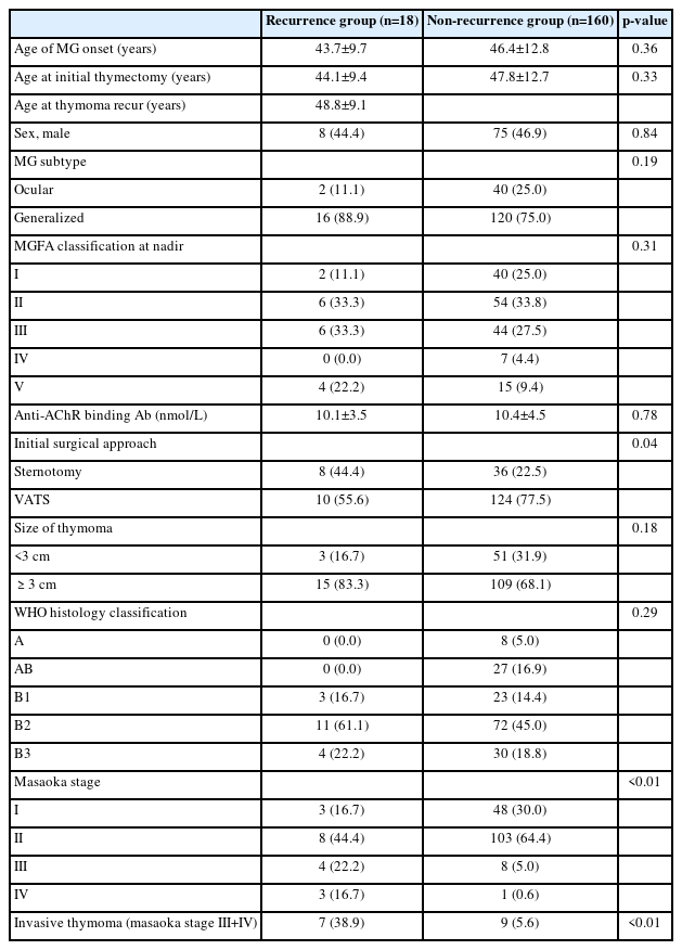
A total of 178 patients with TAMG who were histologically diagnosed with thymoma in Severance Hospital were included. Of these patients, 18 patients (10.1%) experienced recurrence of thymoma and were classified as recurrence group. Mean age at MG onset of the patients in recurrence group was 43.7±9.7 years. Mean age at initial thymectomy and at thymoma recurrence was 44.1±9.4 years and 48.8±9.1 years, respectively. Duration from initial thymectomy to recurrence of thymoma was 56.3±35.3 months. At the point of initial thymectomy, two patients (11.1%) were classified as ocular and 16 patients (88.9%) as generalized MG. Worst MGA classification before initial thymectomy was class I in two patients (11.1%), class II in six (33.3%), class III in six (33.3%), and class V in four patients (22.2%). Median MG ADL score at the point of initial thymectomy was 4.0 (interquartile range [Q1-Q3], 2.3-8.8). The initial World Health Organization (WHO) histology classification was B1 in 16.7%, B2 in 61.1%, and B3 in 22.2%. Masaoka stage II was the most common (44.4%), followed by stage III (22.2%), stage I (16.7%) and stage IV (16.7%). When clinical and histological characteristic were compared between the recurrence and non-recurrence groups (Table 1), no significant differences were observed between groups in the aspect of age of MG onset, age at thymectomy, sex, MGA classification at initial thymectomy, WHO histology classification, and titer of AChR Ab. However, the proportion of patients with invasive thymoma were significantly higher in recurrence group (38.9%) compared to non-recurrence group (5.6%; p<0.01).
State of MG at the point when recurrence of thymoma was detected
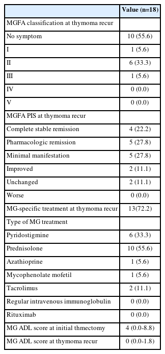
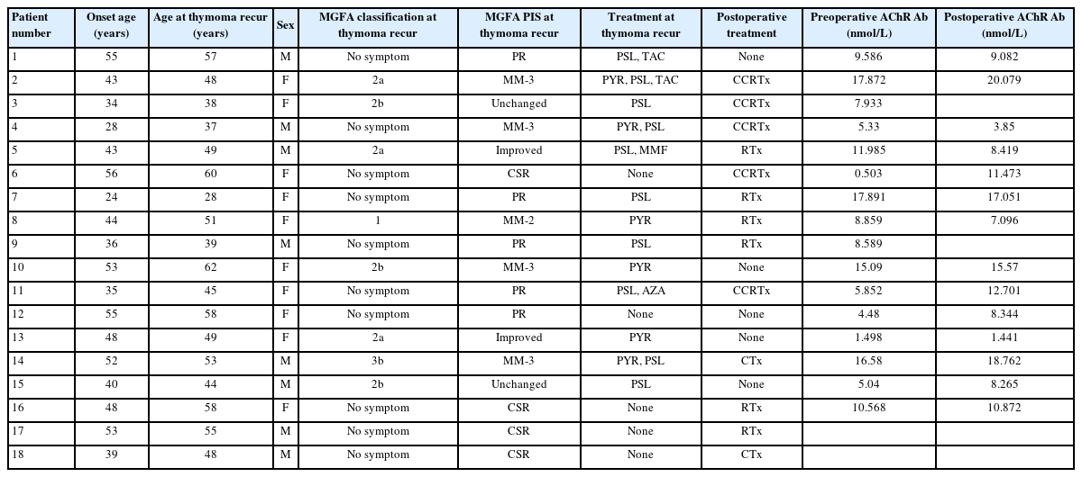
Table 2 presents the clinical and treatment status of the patients with TAMG at the point that thymoma recurrence was radiologically observed. MGA PIS of the patient was complete stable remission in four (22.2%), pharmacologic remission in five (27.8%), minimal manifestation in five (27.8%), improved in two (11.1%), and unchanged in two patients (11.1%). None of the patients were experiencing worsening or exacerbation of MG at this point. In terms of treatment, 10 patients (55.6%) were taking prednisolone and four (22.2%) were on oral immunosuppressants. Five patients (27.8%) were not receiving MG-specific therapy. Median MG ADL score was 0.0 (Q1-Q3, 0.0-1.8). Table 3 presents the detailed clinical characteristics of the 18 patients in recurrence group. In patient 17 and patient 18, recurrence of thymoma was confirmed exclusively on CT imaging without surgical intervention. Of the 14 patients whose serum level of AChR Ab was assessed both before and after the first thymectomy, level of AChR Ab increased in eight patients (57.1%) and decreased in six patients (42.9%).

Clinical status and treatments status of the patients with thymoma-associated myasthenia gravis (MG) at the point of thymoma recurrence
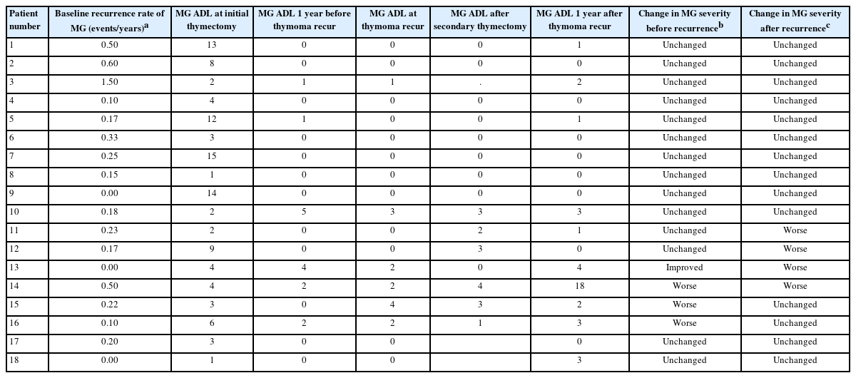
Change of MG ADL before and after the thymoma recurrence
Table 4 displays the baseline recurrence rate of MG and changes in MG ADL score before and after thymoma recurrence. Baseline recurrence rate, calculated by dividing the number of total events of worsening by the total follow-up duration, was 0.29 per years. A total of seven events of worsening occurred during the 2-year period around the recurrence of thymoma, with the worsening rate of 0.19 per year. MG ADL scores were recorded at four time points: 1 year prior to thymoma recurrence, at the time when thymoma recurrence was first detected, following secondary thymectomy, and 1 year after thymoma recurrence. Of the 18 patients in recurrence group, 12 patients remained unchanged during the 2-year period around the thymoma recurrence. Three patients experienced worsening of MG during the 1-year period prior to the recurrence date. In detail, symptoms of MG in patient 14 worsened along with pulmonary tuberculosis infection. Patients 15 and 16 experienced worsening of MG after the self-discontinuation of prednisolone treatment. our patients (patients 11, 12, 13, and 14) exhibited worsening of MG symptoms during the 1-year period after the recurrence date. In these four patients, symptoms of MG worsened 2-4 months after second thymectomy.
Discussion
Of the patients with TAMG who underwent thymectomy, 10.1% experienced recurrence of thymoma during the mean follow-up duration of 10.8 years. Clinical characteristics of MG were not significantly different between the patients who experienced and did not experience recurrence of thymoma. MG was in stable stats in all patients at the time when the recurrence of thymoma was first detected. Of the 18 patients with thymoma recurrence, three and four patients experienced worsening of MG during the 1-year period before and after the recurrence of thymoma, respectively. Rate of MG worsening was 0.19 per year during the 2-year period around the thymoma recurrence. The rate of recurrence was comparable to the baseline rate of worsening during the period other than peri-recurrence period. Patients who experienced worsening of MG around the point of thymoma recurrence had other possible cause of MG worsening besides thymoma recurrence including infection, discontinuation of MG medication and surgery requiring general anesthesia.
In the present study, the recurrence rate of thymoma among the patients with TAMG was 10.1%. This is consistent with the Japanese Association for Research on Thymus data, where 14.8% of patients with thymoma experienced recurrence.4 In other studies, recurrence rates were similarly reported, including 13.7% in a cohort of 95 patients, 14.2% among 148 patients, and 12.3% in a separate analysis, with a median recurrence-free period of 8.2 years, demonstrating comparable outcomes across various populations.2,8,9 In previous studies, risk of thymoma recurrence was significantly higher in patients with early-onset MG, suggesting a potential correlation between earlier onset of the disease and increased recurrence risk.10 Similarly, although not clinically significant, age of onset was younger in recurrence group compared to those in non-recurrence group. In our study, patients with recurrence had a larger tumor size and higher Masaoka stage, which are the known risk factors associated with the recurrence of thymoma.9,11 Consistent with the results from Liu et al.,9 which found that MG symptoms did not significantly influence thymoma recurrence, there were no significant difference in clinical feature of MG between the recurrence and non-recurrence groups in this study.
The precise mechanisms underlying MG exacerbation after thymoma recurrence remain unclear. A previous studies demonstrated that worsening of MG could be a sign of thymoma recurrences.2,6 Especially, Haniuda et al.6 stated that recurrences of thymoma was noticed by the worsening of MG in patients with TAMG. In general, destruction of thymic microenvironment by thymoma and release of nontolerogenic T cells to peripheral circulation are considered to be the main pathomechanism of TAMG.12 However, further studies are required to elucidate whether the recurrent thymoma can also trigger these pathways and result in worsening of MG, or whether the worsening of MG is an indirect result of thymoma recurrence. In addition, in the present analysis, MG exacerbations were associated with respiratory infections, changes in medication, and surgical interventions, which are the known risk factors for the worsening of MG.13,14 Thus, conventionally recognized risk factors for MG exacerbation including infections, emotional stress, microaspiration, medication adjustments, surgery, or trauma, should also be considered in interpreting the cause of MG worsening around the recurrence of thymoma.
This study has several limitations that should be noted. irst, its retrospective design and relatively small sample size may introduce selection bias, limiting the statistical power and generalizability of the findings. This design also constrains our ability to control for potential confounding factors, which may independently influence patient outcomes. Second, clinical decisions on surgical approaches and MG management were individualized, introducing variability in treatment strategies. Third, the exact time of thymoma recurrence cannot be precisely determined, and the time of thymoma recurrence could only be estimated as the time when thymoma recurrence was first observed on chest CT. Lastly, as this is a single-center study, the findings may not fully represent broader populations. Larger, multicenter prospective studies are needed to confirm these observations.
In conclusion, 16.7% and 22.2% of the patients with thymoma recurrence experienced worsening of MG during the year prior to and after the recurrence of thymoma, respectively. However, the rate of worsening was comparable to the generally known rate of MG worsening. In addition, symptom exacerbation in these cases could be attributable to external factors such as infection, discontinuation of medication, or surgical intervention, rather than the recurrence of thymoma itself. These highlights the need for a careful assessment of other contributing factors when evaluating MG worsening in the context of thymoma recurrence.
Supplementary Material
Supplementary Figure 1.
A schematic illustration of recurrence date and 1-year period before and after recurrence date. The change in MG ADL score before and after thymoma recurrence was assessed at the red points in the figure, with patients' MG status evaluated over the 2-year period around the recurrence. MG, myasthenia gravis; ADL, activities of daily living.
Supplementary Figure 2.
A schematic illustration of the periods that were used to calculate the baseline recurrence rate of MG and recurrence rate of MG around thymoma recurrence. The baseline recurrence rate is defined as the number of MG aggravation episodes per year during the period shown in a*. MG, myasthenia gravis. a*Baseline MG follow up duration. b*2-year period around the point of thymoma recurrence.
Acknowledgements
None.
Notes
Author Contributions
Conceptualization: SWK. Data curation: BCO. Formal analysis: BCO, SWK. Investigation: BCO. Methodology: BCO, SWK. Supervision: HYS, YHY, SWK. Visualization: BCO. Writing—original draft: BCO, YHY, SWK. Writing—review & editing: all authors.
Conflicts of Interest
The authors have no potential conflicts of interest to disclose.
Funding Statement
This work was supported by a National Research Foundation of Korea grant funded by the Korean government (Ministry of Science, ICT and Future Planning; 2019R1C1C1009875).
Data Availability Statement
No data are available.
Ethical Approval
This study was approved by the institutional review board of the Severance Hospital (IRB No. 4–2024-1322) with a waiver of informed consent.
Patient Consent for Publication
Not applicable.


